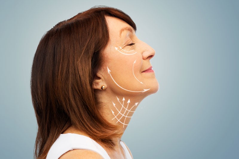
Fat Transplants - Olney, MD
Fat Transplants Olney, MD
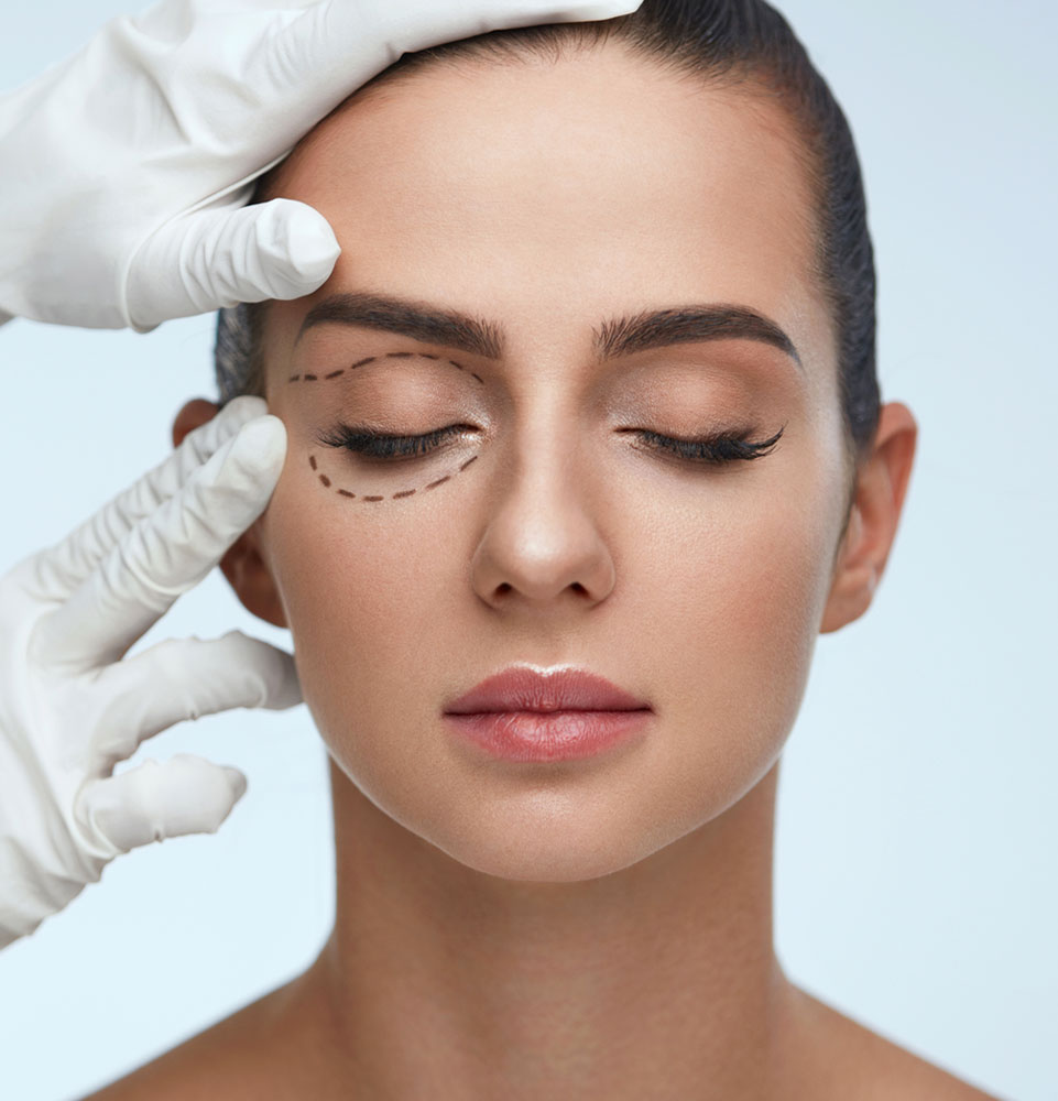
Fat Transplants Olney, MD
Facial deformity due to buccal fat pad loss is a rare but significant complication of dentoalveolar surgery, such as sinus augmentation. We present a case that was referred for correction of facial soft tissue asymmetry in the left submalar region of unknown etiology. The patient, however, states that the soft tissue depression happened following a sinus augmentation procedure that was done by another surgeon. She denied having any trauma or other dental/ facial-related procedure to that area. This asymmetry happened possibly secondary to loss of a section of the buccal fat pad. Whether this was related to the sinus lift procedure could not be ascertained by our investigation. To treat this asymmetry, purified abdominal fat graft was used in multiple small volume injections. The three-year follow-up showed no relapse of the soft tissue facial defect. Fat grafting to correct facial deformity is a safe and reliable treatment with few complications that can replace more invasive surgical procedures.
Buccal fat pad (BFP) is an encapsulated, well-vascularized fatty tissue that consists of a body and four processes. The body has three distinct lobes—anterior, intermediate and posterior—which are located behind the zygomatic arch. There are four processes that extend from the body to buccal space, deep infra-orbital space, pterygomandibular space, and infra-temporal fossa.1 Buccal fat pad functions as a deep and superficial facial space filler, a gliding path for adjacent muscle movement and a cushion to surrounding neurovascular structures during facial and masticatory muscles extension and contractions.1,2,3 Dentoalveolar surgeries, such as upper 3rd molar extraction or sinus augmentation for dental implants, can cause unwanted exposure or release of BFP. The buccal extension of the BFP is the most superficial segment and contains roughly one-third of the BFP volume. This is the main part of BFP, which may become exposed unwantedly during dentoalveolar surgeries, or on purpose for facial recontouring by reducing buccal fullness.3 Over the past several decades, there has been a number of studies that have shown the use of BFT to correct oral and facial deformities by means of extension pertaining to a part of BFP as a pedicle flap to close a defect, or removal of specific lobe(s)/extension of BFP to recontour the face.3-6 This signifies the importance of BFP as facial filler to maintain facial contour and the devastating consequence of its loss on facial appearance. Fat grafting or fat transplant is the removal of excess fat from a donor site with the intention of re-implanting the fat graft in the defect area. It can be used as filler around the lips, nasolabial fold, periorbital area, cheek and chin. It has been shown to successfully correct cosmetic deformity due to trauma, acne, facial hemiatrophy, lipoatrophy, etc.7,8 Autogenous fat transplant was first documented in 1893 by Neuber, who used it to correct facial deformity due to tuberculosis, and then in 1911 by Bruning, who used fat transplant to correct facial asymmetry post-rhinoplasty.9,10 In 1985, Fournier introduced a new harvest technique to the existing liposuction method by aspiration of fat with low-pressure syringe. He called it “ LipoSculpture” and showed that the viability and integrity of adipose tissue is much better preserved with this technique.11 It was not until the 1990s that Coleman added centrifugation to the previous fat liposuction technique, a new fat transfer technique that he described in a series of publications and trademarked as “LipoStructure” for the successful harvest, transport and transplantation of fatty tissue. Coleman centrifuged the harvested fat tissue and injected a specific volume of purified fat, as needed for the defect, in a small volume “parcel of fat” in repeated injections. This technique helps to place the desired volume of fat in a condensed small volume, while keeping the “fat parcels” as separate as possible from each other to provide maximal space between each injected fat tissue. This will help the recipient site to re-vascularize the fat tissue by optimal exposure of graft to blood flow and, hence, survival and regeneration of the transplanted tissue.12,13 In 1994, Carpaneta and Ribeiro showed that smaller injected fat volume, less than 3 mm, had the most survival rate, which confirmed the results of Coleman regarding his fat transfer technique. 14,15 Today, literature shows surgeons using the same main principles as set forth by Coleman, but different steps and procedures have been introduced with successful outcomes and are being used worldwide depending on the surgeon’s experience and comfort level.
Case Report
A 42-year-old woman with no past medical history presented complaining that her face had sunken in after her sinus lift procedure nine months earlier in preparation for dental implants in her left posterior maxilla by another surgeon. She reported being grossly symmetric prior to the sinus augmentation. Upon presentation, the patient was grossly asymmetric, with a volume deficit in the submalar area on the left side without any functional deficit (Figure 1). Intraorally, the patient had three dental implants in the left molar area post sinus lift.

Procedure
The patient was marked and placed in the supine position. 200 cc of tumescent fluid was infiltrated in the lower abdomen (500 cc 0.9% NaCl, 0.5 cc 1:1000 epi, 20 cc 2% lidocaine) and allowed to sit for 30 minutes (Figure 2). A 2.4 mm Tulip was used with a 60 cc syringe at 20 psi pressure to harvest 100 cc of aspirate. The aspirate was transferred into Figure 1. Initial presentation. A. Left lateral view. B. Frontal view. C Right lateral view. A B C Figure 2. Abdominal fat harvest site. 26 JANUARY 2018 • The New York State Dental Journal 20 cc syringes and centrifuged for 45 seconds at 3,000 rpm. The top fatty layer was saved; the fluid on the bottom was discarded. A small oil layer was removed and discarded from the top of the supernatant. The procedure continued with sterile transfer of the fat to a 20 cc syringe. A 19-gauge needle was used to create two points of entry around the left buccal fat pad (Figure 3). One was made along the lateral portion of the zygomatic arch, another in the nasolabial fold. Approximately 15 cc of fat was injected in multiple layers in a fan-like pattern. A portion was injected along the periosteum, another in the muscular layer and a small portion in a most superficial pattern. The left malar area was then massaged to assure even distribution. The defect was slightly overfilled in comparison to the opposite side to balance the initial fat reabsorption.


One Week Post-op
Postoperatively, the patient remained without any complications from the surgery. Her three-year follow-up appointment showed a successful outcome without any visible prolapse
Discussion
Autologous fat tissue has distinctive characteristics, which makes it the material of choice for correcting facial defects. Adipose tissue is readily available, easy to harvest, without rejection failure, non-allergenic and inexpensive. Several histological studies have proven fat graft viability post transplantation. It has been shown that inflammatory cells infiltrate around the graft on the first day. By the fourth day, small vessels surround and penetrate the fat tissue. A small volume (3 mm) of injected fat graft facilitates the angiogenesis process. Over the next several months, the inflammatory cells (lymphocytes, histiocytes, etc) shrink in number, and preadipocytes and new adipocytes will pre-dominate the graft pad. It has been demonstrated histologically that over time, viable adipocytes, fibroblasts, fibrous tissue co-exist at the graft site. This indicates that the clinical results of fat grafting are the interplay between fat tissue survival and regeneration, along with host response fibrosis.16,17 There are a number of anecdotal studies regarding the longevity of fat graft. Some have shown long-term maximal resorption, while some have shown minimal resorption with great long-term results.17 Horl et al. showed there is a 49% loss in the first three months, up to a 55% loss at six months and then minimal loss between 6 and 12 months post fat graft.18 This is the reason for slight overcorrection by many surgeons to overcome this initial fat tissue loss. Current consensus seems to be 20% to 30% volume overcorrection is needed to counter the initial loss while preventing the risk of overcorrection, which is more complicated to reverse.19,20 Fat grafting is a meticulous, multi-step process. The proper way to perform each step is the subject of debate. For example, there is no consensus on the speed or length of fat centrifugation. In the Figure 3. A. Surgical field marking. B. Fat transfer to defect. Note 60cc syringe used for demonstration only. 1cc syringe used for actual procedure. C. Immediate postop. Figure 4. A Left lateral view one-week postop. B. Up-down vertical view one-week postop. C. Frontal view one-week postop. Colman technique, centrifugation is at 3000 RPM for three minutes, while other studies have shown that the viability of fat cells decreases at the high speed of 3000 RPM. Current recommendations are centrifugation at 1000 RPM for two minutes or 1300 RPM for five minutes. 20,21 However, this author’s experience shows centrifugation at 3000 RPM for 45 seconds will have optimal results. Another example would be selecting a donor site for fat harvest. It has been shown that adipocytes can have different size and lipogenic activity depending on the location and function of adipocyte in that site. Hudson et al. showed that fat cells from the outer thigh and buttock have the highest lipogenic activity and are the largest.22 Subsequently, a number of publications showed lower resorption rate and higher fat survival when the fat is grafted from the outer thigh or buttock area. However, later on, two separate studies by Rohrich et al. and Ullman et al. showed no significant statistical difference in fat transplant outcome harvested from any available donor sites, including the abdomen, outer thigh, knee, buttock, flank and breast.23,24 Today, donor sites are selected by the amount of fat availability, patient preference and surgeon comfort. Complications from fat graft are rare and mostly mild in nature, but they can be significant and cause devastating morbidity. The most common reported complication is overcorrection, especially in the infraorbital area.24-26 Other common, non-significant complications include postop edema, bruising, bleeding and tenderness. Some of the more significant complications reported are formation of small fat cyst, fat hypertrophy, fat necrosis and infections, but these complications are rare.
See The Results For Yourself

Conclusion
Fat grafting to correct facial deformity is a safe and reliable treatment with few complications. A review of the literature shows great outcomes with fat grafting to treat facial deformities. The transplanted fat undergoes revascularization and regeneration, which becomes integrated into the defect area. Fat grafting gives a natural shape or contour to the area, which transitions smoothly to adjacent structures. In the case reported here, we had successful results with complete patient satisfaction three years post-facial fat grafting. ???? Queries about this article can be sent to Dr. Tebyanian at anistebyanian@gmail.com.
References
- Zhang HM, Yan YP, Qi KM, Wang JQ, Liu ZF. Anatomical structure of the buccal fat pad and its clinical adaptations. Plast Reconstr Surg 2002 Jun;109(7):2509-18; discussion 2519-20.
- Stuzin JM, Wagstrom L, Kawamoto HK, Baker TJ, Wolfe SA. The anatomy and clinical applications of the buccal fat pad. Plast Reconstr Surg 1990 Jan 85(1):29-37.
- Matarasso A. Buccal fat pad excision: aesthetic improvement of the midface. Ann Plast Surg 1991 May 26(5):413-8.
- Khiabani K, Keyhan SO, Varedi P, Hemmat S, Razmdideh R, Hoseini E. Buccal fat pad lifting an alternative open technique for malar augmentation. J Oral Maxillofac Surg (2013), doi: 10.1016/j.joms.2013.10.002.
- Hanazawa Y, Itoh K, Mabashi T, Sato K. Closure of oroantral communications using a pedicled buccal fat pad graft. J Oral Maxillofac Surg 1995 Jul 53(7):771-5 discussion 775-6.
- Baumann A, Ewers R. Application of the buccal fat pad in oral reconstruction. J Oral Maxillofac Surg 2000 Apr 58(4):389-92 discussion 392-3.
- Donofrio LM. Techniques in facial fat grafting. Aesthet Surg J 2008 Nov-Dec 28(6):681-7. doi: 10.1016/j.asj.2008.09.003.
- Xie Y, Li Q, Zheng D, Lei H, Pu LL. Correction of hemifacial atrophy with autologous fat transplantation. Ann Plast Surg 2007 Dec 59(6):645-53.
- Neuber F. Fat transplantation. Chir Kongr Verhandl Dsch Gesellch Chir 1893 20:66.
- Bruning. Contibution a l&rsquo etude des greffes adipeuses. Bull Acead R Med Belgique 1914 28:440-430.
- Fournier P. Microlipoextraction et microlipoinjection. Rev Chir Esthe´ t lang Fr 1985 10:36e40.
- Coleman SR. Facial recontouring with lipostructure. Clin Plast Surg 1997 Apr 24(2):347-67.
- Coleman SR. Structural fat grafts: the ideal filler? Clin Plast Surg 2001 Jan 28(1):111-9.
- Carpaneda CA, Ribeiro MT. Study of the histological alterations and viability of the adipose graft in humans. Aesthetic Plast Surg 1993 17:43&ndash 7.
- Carpaneta C. Percentage of graft viability versus injection volume in adipose auto transplant. Adv Plast Surg 1994 18:16&ndash 9.
- Sclafani A, Romo T III, Parker A, Cormick S, Cocker R, Jakono A. Autologous collagen dispersion (Autologen) as a dermal filler. Clinical observations and histologic findings.Arch Facial Plast Surg 2000 2:48.
- Sommer B, Sattler G. Current concepts of fat graft survival: histology of aspirated adipose tissue and review of the literature. Dermatol Surg 2000 26:1159&ndash 66.
- Horl HW, Feller AM, Biemer E. Technique for liposuction fat reimplantation and long-term volume evaluation by magnetic resonance imaging. Ann Plast Surg 1991 26:248.
- Fulton JE, Parastouk N. Fat grafting. Dermatol Clin 2001 Jul 19(3):523-30.
- Xie Y, Zheng DN, Li QF, Gu B, Liu K, Shen GX, Pu LL. An integrated fat grafting technique for cosmetic facial contouring. J Plast Reconstr Aesthet Surg 2010 Feb 63(2):270-6. doi: 10.1016/j.bjps.2008.11.016. Epub 2008 Dec 13.
- Ferraro GA, De Francesco F, Tirino V, Cataldo C, Rossano F, Nicoletti G, D&rsquo Andrea F. Effects of a new centrifugation method on adipose cell viability for autologous fat grafting. Aesthetic Plast Surg 2011 Jun 35(3):341-8. doi: 10.1007/s00266-010-9613-8. Epub 2010 Nov 11.
- Hudson DA, Lambert EV, Block CE. Site selection for autotransplantation: some observations. Aesthetic Plast Surg 1990 14:195.
- Rohrich RJ, Sorokin ES, Brown SA. In search of improved fat transfer viability: a quantitative analysis of the role of centrifugation and harvest site. Plast Reconstr Surg 2004 113:391&ndash 5.
- Ullman Y, Hyams M, Ramon Y, et al. Enhancing the survival of aspirated human fat injected into nude mice. Plast Reconstr Surg 1998 101:1940&ndash 4.
- Donofrio L. Technique of periorbital lipoaugmentation. Dermatol Surg 2003 29:92&ndash 8.
- Fulton JE, Suarez M, Silverton K, Barnes T. Small volume fat transfer. Dermatol Surg 1998 24:857e65.
- Miller J, Popp J. Fat hypertrophy after autologous fat transfer. Ophthalmic Plast Reconstr Surg 2002 18:228&ndash 31.
- Donofrio LM. Techniques in facial fat grafting. Aesthet Surg J 2008 Nov-Dec 28(6):681-7. doi: 10.1016/j.asj.2008.09.003.